|
| Guess What! - ESTP Case 21 |
The lesion was observed in an approximately 4 year old male cynomolgus monkey (Macaca fascicularis) from a subchronic toxicity study with daily subcutaneous application. No clinical finding was reported relating to the lesion and there were no gross observations in this animal at necropsy.
| Click on the images below for a larger view. |
|
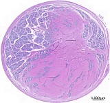
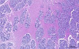
Fig. 1: H&E
|
|
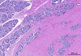
Fig. 2: H&E
|
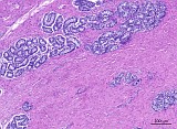
Fig. 3: H&E
|
|
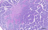
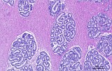
Fig. 4: H&E
|
|
Fig. 5: H&E
|
Fig. 6: H&E
|
Morphologic Description
Case 21 from an approximately 4-year old male cynomolgus monkey (Macaca fascicularis) shows a bilateral replacement of the normal lobular testicular parenchyma in transverse sections of two testes (figs.1 and 4). Variable amounts of mature collagen connective tissue were obvious separating and displacing the immature seminiferous tubules.
The testis shown in figs. 1-3 was markedly affected while the contralateral one (figs. 4-6) showed only slight changes. At higher magnification different areas of the lesion are visible. Fig. 3 and 6 show islands of remaining immature/inactive seminiferous tubules. Active spermatogenesis is not present.
Proposed Diagnosis
Testicular fibrous hypoplasia
| Click on the image below for a larger view. |
|
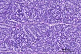
Fig. 7: H&E
|
Discussion
Due to variable age/maturity of cynomolgus monkeys in toxicological studies, differences in size and weight of testes are common. Smaller testes mostly relate to immature/pubertal or inactive testicular tissue with regular lobular architecture but without active spermatogenesis (fig. 7). Immaturity can be confirmed by investigation of epiphyseal closure of the femoral bone. The case shown here was found in a study in which two still juvenile animals showed a bilateral fibrous replacement of varying amounts in both testicles.
Testicular fibrous hypoplasia in cynomolgus monkeys is reported as uni- or bilateral background lesion (1, 2). It is assumed to be a congenital condition that is more prevalent in recent years and apparently associated with the importation of cynomolgus monkeys from Indo-China (1).
Nine diagnoses were received for this case:
Three diagnoses referred to fibroadenoma of the mammary gland, multifocal intestinal carcinoid tumor or adenocarcinoma with smooth muscle proliferation originating from gastrointestinal or urogenital tract, but can be excluded as the present lesion developed in the testis.
Six contributing participants recognized the testes as affected organs. By these contributors an increase of interstitial stroma or fibrous tissue was consistently diagnosed. It was attributed to interstitial fibrosis, hypoplasia or immaturity. A developmental anomaly or cryptorchism was proposed to be the underlying cause. One sender presumed it to be normal for the species.
As differential diagnoses, fibroma and intratesticular fibromatosis were proposed.
Morphologically, the replacing tissue was mature dense connective tissue. The origin of the lesion may be the interstitial connective tissue of the testis, or the peritubular cells which are transformed in human testicular fibrosis (3).
Fibrosis as result of a tissue injury/trauma as well as fibroma may be considered as differentials.
Benign fibroblastic proliferations are mostly considered as reactive, nonneoplastic and of fibroinflammatory origin. They encompass lesions with morphology of cellular pseudosarcomatous to fibrotic hypocellular proliferations which mostly appear nodular and may contain calcification or bone formation (4). Attributed to their variable characters, they are called fibrous pseudotumors, fibromatous tumors and tumor-like lesions (4, 5) or fibromatous lesions (6).
The term fibroma has been used for a variety of benign fibroblastic proliferations involving the testicular parenchyma, the tissue layers enclosing the testicles (testicular tunics), and other paratesticular sites. Attributed to the origin of the neoplasias arising from gonadal stroma and the homologous appearance to ovarian fibromas, the term "fibroma of gonadal stromal origin" was proposed by Jones et al. in humans. They could be well distinguished from sex cord-stromal tumors which may have prominent fibromatous components but also have sex cord components (4).
The present lesion is not regarded as a tumor since well differentiated cells do not show any pleomorphism, mitoses or variations in activity. Moreover, the growth pattern is regular without an infiltrative or nodular growth as described for most tumors. Due to the juvenile status of the animals we propose a developmental anomaly, because there is no evidence of other causes, although another etiology cannot be excluded. An influence on Leydig cell activity or a trophic impairment of the seminiferous tubules may be a possible underlying cause of associated lack of active spermatogenesis. Leydig cells in non-human primates and men occur singly or may be arranged in rows or clumps of varying size within a very loose connective tissue (7). Since there are no further morphological differences, especially regarding Leydig cells, compared to other juvenile testes in this cohort, we did not take an influence on Leydig cells into consideration as the underlying cause of the observed lesion.
Because of the existing description of this lesion in the testes in cynomolgus monkeys (1, 2), we choose the term "fibrous hypoplasia". However, other terms such as interstitial fibrosis or increased stromal tissue may be also appropriate.
References
- Creasy D (2012) Reproduction of the Rat, Mouse, Dog, Non-human Primate and Minipig. In: McInnes EF (ed.) Background Lesions in Laboratory Animals: A Color Atlas. Saunders Elsevier Edinburgh, London, New York, 109-110.
- Patrick DJ, Rebelatto MC (2015) Toxicologic Pathology and Background Lesions of Nonhuman Primates. In: Bluemel J, Korte S, Schenck E, Weinbauer G (eds.)The Nonhuman Primate in Nonclinical Drug Development and Safety Assessment. Elsevier London, San Diego, Waltham, Oxford, 252.
- Mayerhofer A (2013) Human testicular peritubular cells: more than meets the eye. Reproduction 145: 107-116.
- Jones M, Young R, Scully R (1997) Benign Fibromatous Tumors of the Testis and Paratesticular Region: A Report of 9 Cases with a Proposed Classification of Fibromatous Tumors and Tumor-Like Lesions. Am J Surg Pathol 21: 296-305.
- Ugras S, Yesil C (2009) Fibrous pseudotumors of tunica albuginea, tunica vaginalis and epididymis: Report of two cases. Cancer Epidem 33: 69-71.
- White M, Hilsenbeck J, Waters WB (2006) Fibromatous Periorchits of Testes. Urology 67(3): 623.e15-16.
- Fawcett DW, Neaves WB, Flores MN (1973) Comparative Observations on Intertubular Lymphatics and the Organization of the Interstitial Tissue of the Mammalian Testis. Biol Reprod 9: 500-532.
|
|