|
| Guess What! - ESTP Case 13 |
Animal / species: Rattus norvegicus, Wistar Rat, Rj:WI (IOPS HAN).
Sex / age / weight : Female animal, 742 Days of age, terminal body weight 365g.
Use in toxicology: Animal in a standard carcinogenicity study, control group.
Spontaneous finding: No macroscopic finding.
Slide: Section from the thymus, standard paraffin embedding, H&E stained slide.
| Click on the images below for a larger view. |
|
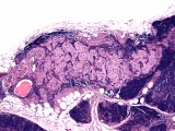
Fig. 1: H&E, x5
|
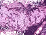
Fig. 2: H&E, x10
|
|
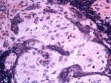
Fig. 3: H&E, x40
|
Morphologic Description
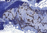
Case # 13 showed the histopathological appearance of an atrophic thymus (normal involution, age-related) with a mixture of hyperplastic epithelial cords and an uncommon stromal proliferation. The lesion appears well circumscribed amongst the thymic parenchyma.
Proposed Diagnosis
Benign schwannoma
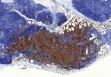
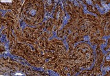
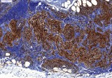
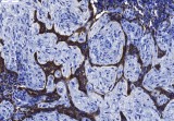
There is a well circumscribed, unencapsulated proliferative stromal mass embedded within an atrophic thymic lobule (Fig. 1). The mass does not infiltrate peripheral thymic lobules. It is mainly composed of fusiform to ovoid neoplastic cells (Figs. 2-3). The nuclei are ovoid to fusiform with vesicular chromatin and occasional single nucleoli. The cells have abundant amphophilic to myxoid cytoplasm with indistinct cell borders (Fig. 3). Anisokaryosis and anisocytosis are minimal and the mitotic index is low. Bundles of neoplastic fusiform cells are S-100 (Figs. 4-5) and vimentin positive (Fig. 6), separated by small clusters of cytokeratin positive cells (hyperplastic epithelial cords) (Figs. 7-8). Immunohistochemical results favor the diagnosis of a benign schwannoma associated with cords of hyperplastic thymic epithelium. Epithelial hyperplasia is commonly observed in aging Wistar rats.
| Click on the images below for a larger view. |
|
Fig. 4: S-100, x11
|
Fig. 5: S-100, x40
|
|
Fig. 6: vimentin, x20
|
|
Fig. 7: cytokeratin, x20
|
Fig. 8: cytokeratin, x40
|
Discussion
Two contributors diagnosed a benign schwannoma. Three out of 13 contributors mentioned epithelial cell hyperplasia. Another one specified "clusters of epithelial cells" which are part of the lesion described above (hyperplastic epithelial cords). There were three participants suggesting sinusoidal dilatation, angiomatous lesion or vascular leiomyoma. However, the proliferation of fusiform, S-100 positive cells with a large, indistinct cytoplasm is indicative of a neurogenic rather than a vascular origin.
One contributor diagnosed a carcinoid or an ectopic C-cell tumor. Endocrine or neuroendocrine tumors have generally clusters of round to ovoid epithelial cell amongst a regular fibro-vascular stroma. Their cytoplasm is distinct with an eosinophilic granular appearance; the nucleus is round, located centrally or slightly eccentric. In the case presented the microscopic appearance is slightly different from a classical endocrine or neuroendocrine tumor.
Two contributors diagnosed a benign thymoma. Thymoma corresponds to primary involvement of thymic epithelial cells often admixed with lymphocytes. Various levels of differentiation may be present ranging from tumors with a predominantly normal thymic structure and medullary differentiation to thymomas composed of a mixture of epithelial cells and lymphocytes lacking medullary differentiation. Furthermore, thymomas can be categorized as epithelial, neuroendocrine, spindeloid or myoid type.
The present case might be diagnosed as a spindeloid type thymoma but the fusiform part is vimentin and S-100 positive and thus excluding a thymic epithelial cell population.
Apart from thymomas or thymic lymphosarcomas, other primary neoplasms of the thymus are extremely rare. Neoplasms like paraganglioma or lipoma are very uncommon and may originate in tissues adjacent to the thymus.
The innervation of the thymus is described in the literature and appears to be complex. In the rat, catecholaminergic and cholinergic nerve fibers are detected along the vessels and in the thymic parenchyma in specific locations. They are linked with the parasympathetic system and the phrenic nerve, and play a key role in the homeostasis of neuroimmune modulation. In the subcapsular region of the thymus, the parenchymal cholinergic fibers originate exclusively from phrenic nerve branching. However, no somatic phrenic nerve branching is detected in any part of the thymus.
In the mice the thymus is innervated by acetylcholine-esterase-positive fibers of the vagus, the recurrent laryngeal, and the phrenic nerves.
The present benign schwannoma may have arised from this complex and branched intrinsic innervation. This dense nervous network might explain why the lesion seems to be well embedded within the parenchyma without any associated compression.
References
- Boorman et al. (1990) Pathology of the Fisher Rat. 392-393.
- Mignini F, Sabbatini M, D'Andrea V, Cavallotti C (2010) Intrinsic innervation and dopaminergic markers after experimental denervation in rat thymus. Eur J Histochem 54: e17
- Bulloch K, Pomerantz W (1984) Autonomic nervous system innervation of thymic-related lymphoid tissue in wildtype and nude mice. J Comp Neurol 228: 57-68.
|
|