|
| Guess What! - ESTP Case 4 |
The lesions of the uterus were observed as an incidental finding in a
4 y 8 m old Cynomolgus monkey (Macaca fascicularis) control female of
a subchronic toxicity study. Macroscopically, the organ showed no
abnormalities.
| Click on the images below for a larger view. |
|
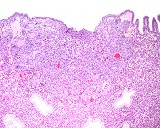
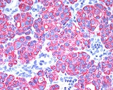
Fig. 1: H&E, x10
|
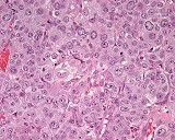
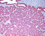
Fig. 2: H&E, x40
|
Morphologic Description
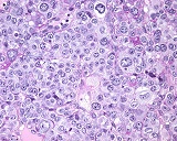
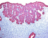
Two epithelial plaques were observed microscopically in the uterus of a 4 years 8 months old non-pregnant cynomolgus monkey. The plaques were located opposite to each other and extended from the columnar epithelium lining the uterine lumen into the functional layer of this organ. Multifocally, the plaques were in direct contact with the uterine lumen and uterine epithelium was lacking at these sites. Both plaques consisted of clusters and nests of epithelial cells, that were separated by a scanty fibrous stroma. The plaque forming cells had large vesicular nuclei with a distinct nucleolus and abundant faintly basophilic cytoplasm. Frequently cells revealed giant nuclei and occasionally also bi-nucleated cells were found scattered among the others. Generally, marked cellular pleomorphism was one of the most prominent features of this lesion. Some cells had vacuoles. PAS-reaction revealed PAS-positive granular material (probably representing glycogen) in several cells. Mitotic figures were often seen in both plaques. In the periphery of the plaques slight infiltration by polymorphonuclear leukocytes could be observed. Plaque cells stained positively for pan-cytokeratin, ck 7, ck 8, ck 18 and ck 19 while they had (in contrast to strongly stained uterine cells) only a weak reaction for vimentin.
Proposed Diagnosis
Spontaneous epithelial plaques of the uterus in a non-pregnant monkey
Differential Diagnoses and Discussion
Adenoma/adenocarcinoma was suggested by some contributors as diagnosis. Although immunohistochemistry clearly identified uterine gland or lining epithelium as source of the cells of these lesions, no glandular structures were observed within the plaques. Decidual reaction/deciduoma was a further diagnosis. However, mesenchymal origin of this lesion (positivity for desmin of cells at least in mice) has been proven, while cells of the two plaques were positive for a number of cytokeratins characteristic of columnar epithelium. Granular cell tumor of the uterus, which was a further differential diagnosis sent in, were described in rats and humans. They normally lack the cellular pleomorphism seen in the epithelial plaques. Furthermore occurrence of PAS-positive granules in the cells are a key feature of these tumors, while they were only identified in part of the cells in the epithelial plaques. S-100 immunohistochemistry was not performed on the plaques. Negative results of this stain would support the diagnosis of the lesions as epithelial plaques and further exclude a diagnosis as granular cell tumor.
Uterine epithelial plaques are a common but not ubiquitous endometrial response of primates to implantation in the early stage of pregnancy. They have not been observed in human beings. Epithelial plaques are transient structures and have been described in rhesus monkeys, baboons, marmosets, green monkeys, dusky leaf monkeys and cynomolgus monkeys. They have been most extensively studied in the rhesus monkey and in the baboon. The epithelial plaque reaction of endometrium has been observed one day after implantation in primary implantation sites of rhesus monkeys. The response begins about one day later at the secondary site of implantation in the rhesus monkey and in the baboon plaque formation is seen at the margin of the implantation site in the first days of implantation. Light microscopically it can be seen that individual cells of the uterine luminal epithelium and the necks of the glands lose their columnar configuration and undergo a great increase in size. The rapid enlargement of the epithelial plaque cells leads to formation of clusters and nests enclosed in a basal lamina. This mass of cells forms a pad around the embryo which is in turn surrounded by a zone of extreme edema. Subsequently, the plaque cells start to store glycogen and show clusters of small irregular granules. Some of the epithelial plaque cells begin to degenerate by day 16 of pregnancy, and the degree of degeneration increases so that by day 30 only a few intact plaque cells are still present. Degeneration of plaque cells is accompanied by a distinct leukocytic invasion of the plaque region. As the degeneration of plaque cells proceeds, macrophages accumulate in this region, leaving a zone free of plaque cells but containing debris and a number of lipid-filled macrophages. This zone persists as discontinuous patches between cytotrophoblast and decidua for a considerable time.
| Click on the images below for a larger view. |
|
Fig. 3: PAS, x40
|
Fig. 4: Pan-cytokeratin immunostaining, x10
|
|
Fig. 5: Pan-cytokeratin immunostaining, x40
|
Fig. 6: Ck 8 immunostaining, x10
|
References
- Scully RE, Poulsen HE (Ed.) Histological Typing of Female Genital Tract Tumours. World Health Organization, International Histological Classification of Tumours, 1994, 2nd ed, Springer-Verlag, Berlin and Heidelberg
- Kaspareit J et al.: Spontaneous epithelial plaques in the uterus of a non-pregnant cynomolgus monkey (Macaca fascicularis). Exp Toxic Pathol 56: 9-12 (in press)
|
|