|
| Guess What! - ESTP Case 7 |
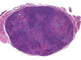
The case is from a 112-week-old female Wistar rat. The left side of the thyroid gland was moderately enlarged, grey-red, and showed a weight of 0.053 g.
| Click on the images below for a larger view. |
|
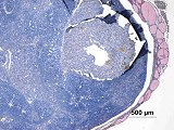
Fig. 1: H&E, x2.5
|
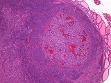
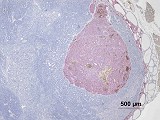
Fig. 2: H&E, x5
|
|
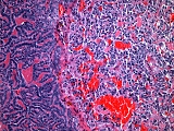
Fig. 3: H&E, x20
|
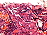
Fig. 4: H&E, x40
|
Morphologic Description
The lesion is characterized by an expansile growing mass, replacing almost the complete thyroid gland. It is surrounded by a thin capsule and consists of bands or ribbon-like structures, intermingled with solid and follicular-like areas. The cells have sparse light eosinophilic cytoplasm with large, dark basophilic staining nuclei. Occasionally the nuclei have a light vesicular appearance. There are focal areas of necrosis and in single areas the capsule seems to be infiltrated by the tumor cells. The proliferation index is moderate with 1-2 mitotic figures per HPF.
Within this tumor located is a smaller mass, which is encapsulated by a well-defined thin capsule and shows compression of the surrounding tissue. The cells are arranged in lobules, separated by a thin fibro-vascular stroma. Within the lesion are numerous blood-filled spaces. The large cells have indistinct cell borders, a moderate amount of light eosinophilic and granular cytoplasm and round to oval pale nuclei. Mitotic figures are very rare (1 observed in the whole tumor).
Proposed Diagnosis
Follicular cell carcinoma and C-cell adenoma
Differential Diagnoses and Discussion
Follicular cell adenoma could be considered as an alternative diagnosis. However, the presence of pleomorphic cells, focal areas of necrosis, the relatively high mitotic index and an indication of capsular infiltration led to the diagnosis of the malignant variant of this tumor.
Other contributors suggested the diagnosis of a tumor originating either from follicular cells, C-cells or parathyroid gland. With the different immunohistochemical staining (large mass: slightly positive for thyroglobulin (Fig. 5), negative for calcitonin (Fig. 6) and parathormone (Fig. 7); small mass: positive for calcitonin, negative for thyroglobulin and parathormon) the different histiogenesis of the tumor cells could be demonstrated. As both masses stained negative for parathormone the parathyroid gland was not considered as origin of one of the both tumors.
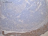
One of the contributing participants suggested the diagnosis Entrapped parathyroid gland. From the H&E stained picture this might have been an alternative diagnosis, but as the immunohistologic stain for calcitonin (Fig. 6) shows a clear positive reaction of the cells of the smaller tumor mass, the diagnosis of a C-cell tumor was made. Also the blood-filled spaces are a typical morphologic feature in these tumors. The malignant variant was not considered, as no evidence of malignancy (capsular infiltration, necrosis, high mitotic index) was observed.
| Click on the images below for a larger view. |
|
Fig. 5: immunohistological stain for thyroglobulin
|
Fig. 6: immunohistological stain for calcitonin
|
|
Fig. 7: immunohistological stain for parathormone
|
References
- Boorman GA, DeLellis RA (1983) C cell adenoma , thyroid, rat. In: Jones TC Mohr U, Hunt RD (eds) Monographs on pathology of laboratory animals. Endocrine system. Springer, Berlin Heidelberg New York Tokyo, pp 197-200
- Capen CC, Martin SL (1989) The effect of xenobiotics on the structure and function of thyroid follicular cells and C-cells. Toxicol Pathol 17: 266-293
- Hardisty JF, Boorman GA (1990) Thyroid gland. In: Boorman GA, Eustis SL, Elwell MR, Montgomery CA, MacKenzie WF (eds) Pathology of the Fisher rat. Reference and Atlas. Academic Press, San Diego New York London, pp 501-536
- Kaspareit-Rittinghausen J, Wiese K, Deerberg F, Nitsche B (1990) Incidence and morphology of spontaneous thyroid tumors in different strains of rats. J Comp Path 102: 421-432
- Pilling AM, Jones SA, Endersby-Wood HJ, McCormack NAM, Turton JA (2007) Expression of thyroglobulin and calcitonin in spontaneous thyroid gland tumors in the Han Wistar rat. Toxicol Pathol 35: 348-355
- Pour PM, Wilson JT, Salmasi S (1983) Adenoma, carcinoma, parathyroid, rat. In: Jones TC Mohr U, Hunt RD (eds) Monographs on pathology of laboratory animals. Endocrine system. Springer, Berlin Heidelberg New York Tokyo, pp 281-287
|
|