|
| Guess What! - ESTP Case 14 |
Cynomolgus Monkey, female, approximately 4 years of age.
At necropsy the left ovary was enlarged.
| Click on the images below for a larger view. |
|
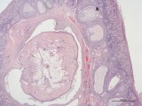
Fig. 1: H&E, x2
|
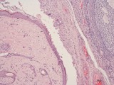
Fig. 2: H&E, x10
|
|
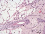
Fig. 3: H&E, x20
|
Morphologic Description
Case 14 showed an unilateral expansile growing mass within the ovary of a four year old purpose bred cynomolgus monkey. The remaining ovarian tissue (follicles, stroma) and the contralateral ovary appeared normal. The bleeding records of the animal revealed normal menstrual cycles. The tumor is well demarcated and lined by keratinized epithelium and contains mature skin adnexa including sebaceous glands and hair shafts as well as mature tissue elements originating from other germ cell layers including adipose tissue, bone, ciliated columnar epithelia interspersed with goblet cells and smooth muscle.
Proposed Diagnosis
Mature ovarian teratoma
Synonyms: Dermoid cyst, benign cystic teratoma
Mature (or cystic) teratomas are by far the most common germ cell tumors in women, accounting for approximately 20% of all human ovarian neoplasms. They have also been described in nonhuman primates including cynomolgus monkeys, rhesus monkeys and domestic animals like bitch, sow, mare and cow as well as in laboratory mice and rats. Mature teratomas may become huge tumors, but are generally considered benign. Rare malignant forms may origin from malignant transformation of any cell type present in the teratoma or are described as "immature teratoma".
Some authors use the term dermoid cyst as a frank synonym for teratoma, others distinguish between dermoid cyst (consisting of ectodermal structures like skin and appendices) and true teratoma (consisting of tissues from all germ cell layers).
Differential Diagnoses and Discussion
28 diagnoses were received for this case. By far the most contributors suggested the diagnosis teratoma or one of its synonyms.
Other contributors suggested dermoid metaplasia or ovarian hamartoma.
The commonly recognized parthenogenetic theory of the origin of ovarian teratomas suggests that they originate from a totipotent primordial cell, and, as they are homozygous likely derived from a single ovum after the first meiotic cell division. In contrast metaplasia describes the reversible transformation of one single cell type into another.
Hamartomas originate from cells, which are physiologically present in the organ affected. Ovarian hamartomas derived from interstitial cells or of vascular origin have been described in animals. They do not include cells from various germ cell layers.
References
- Kaspareit J, Friderichs-Gromoll S, Buse E, Habermann G (2007): Spontaneous neoplasms observed in cynomolgus monkeys (Macaca fascicularis) during a 15-year period. Exp Toxicol Pathol 59:163–9
- Chalifoux LV (1993) In: Jones TC, Mohr U, Hunt RD (eds) Nonhuman Primates II, Ovarian teratoma, Macaca mulatta. Springer Berlin Heidelberg New York Tokyo
- Talerman A (2002) Germ cell tumors of the ovary. In: Kurman RJ (ed) Blaustein's pathology of the female genital tract Springer, Berlin Heidelberg New York Tokyo, pp 967-1033
- Foley GL and Johnson R (1990) A congenital interstitial cell hamartoma of the equine ovary Vet Pathol 27: 364-366
- Schlafer H and Miller RB (2007) In: Jubb, Kennedy and Palmers Pathology of domestic animals. Saunders Elsevier, Edinburgh
|
|