|
| Guess What! - ESTP Case 3 |
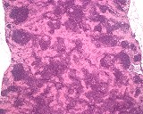
This lesion of a mesenteric lymph node was observed as an incidental finding in a 20-week-old control male CD-1 mouse from a subchronic toxicity study. Macroscopically, the organ showed no abnormality.
| Click on the images below for a larger view. |
|
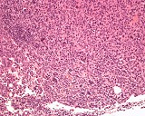
Fig. 1: H&E, x5
|
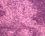
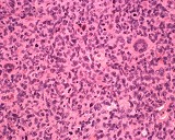
Fig. 2: H&E, x40
|
Morphologic Description
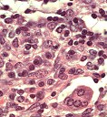
The lesion was composed of histiocytic-like cells with abundant eosinophilic cytoplasm and elongated and partly pleomorphic nuclei and was located mainly in the paracortical regions of the lymph node. The general lymph node architecture was considered normal. Due to their syncytial appearance a more solid growth pattern is suggested. Mitotic figures and inflammatory cells were observed occasionally. The occurrence of giant cells was rare (Fig. 3). Staining with Masson-Goldner showed no increase in collagen fiber formation. Gram and Giemsa staining revealed no presence of any bacteria.
Proposed Diagnosis
Hyperplasia of interdigitating dendritic cells
This lesion is commonly seen in response to a variety of chronic lesions. Hyperplasia of dendritic reticular cells, more specifically interdigitating cells, is seen in response to viral infection, irradiation or other processes. Focal lesions may be noted in the paracortex while extensive lesions involve the entire lymph node. In the present case, the lesion occurred in more than 10 percent of the animals and was found in the mesenteric but also in the mandibular lymph nodes. A latent MHV infection was proofed.
Differential Diagnoses and Discussion
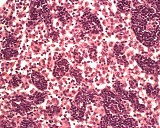
"Sinus histiocytosis": The diagnosis of sinus histiocytosis was proposed by a number of participants. Sinus histiocytosis is frequently present in lymph nodes draining inflammatory lesions. Their cytoplasm has a distinct eosinophilic appearance and may contain phagocytozed material. In general, the histiocytes are regularly formed and no evidence of cellular cohesion or mitotic figures is given (Fig. 4).
"Histiocytic Sarcoma": Early histiocytic sarcoma is similar morphologically to dendritic reticular cell hyperplasia and may allow for difficult interpretation. Normal architecture of the lymph node is distorted. (Figs. 5-6).
| Click on the images below for a larger view. |
|
Fig. 3: H&E, x63
|
Fig. 4: H&E, x40
|
|
Fig. 5: H&E, x20
|
Fig. 6: H&E, x40
|
References
- Frith, CH, Pattengale PK, Ward JM (1985) A color atlas of hematopoietic pathology of mice. Toxicology Pathology Associates, Little Rock, Arkansas
- Ward JM: Classsification of reactive lesions of lymph nodes. In: Monographs on Pathology of Laboratory Animals: Hemopoietic Tissue edited by Jones TC, Ward JM, Mohr U, Hunt RD. Springer Berlin, pp 155-161
|
|