|
| Guess What! - ESTP Case 27 |
The lesion was observed as an incidental finding in an approximately 9-month old female Himalayan rabbit from a reprotoxicity study performed in the late eighties of the last century.
| Click on the images below for a larger view. |
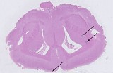
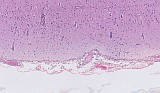
Fig. 1: Overview on a brain section (cerebrum) from a rabbit
with two spots in the right and
one submeningeal spot in the left hemisphere
|
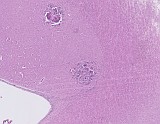
Fig. 2: Higher magnification of the two right spots
|
|
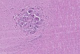
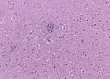
Fig. 3: Higher magnification of the lower spot from Fig. 2
|
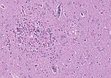
Fig. 4: Higher magnification of the left submeningeal spot
|
|
Fig. 5: Overview on meninges and outer cortex
|
Fig. 6: Higher magnification of the outer cortex
|
Proposed Diagnosis
Inflammation, granulomatous and Infiltrate, inflammatory cell, perivascular ("Encephalitozoonosis")
For Case 27 twelve answers were received, eleven of which described the proposed diagnoses of chronic multifocal, non-suppurative, granulomatous meningoencephalitis with perivascular lymphoid cuffing and glial nodule formation in the cerebrum and meninges of a rabbit. Ten of the participants correctly suspected an infection with Encephalitozoon cuniculi, one respondent thought of a possible viral background. One colleague obviously expected a less simple solution and assumed an infiltration of the cerebrum with a malignant glioblastoma instead.
The case was chosen because it presents features that nowadays are rarely seen in animals from barriered colonies but occurred occasionally some decades ago. The disease still may cause a problem in pet rabbits. Most infections remain rather latent while severely affected animals show neurological symptoms, ranging from occasional shaking of the head to minute-long tilted head and even torticollis. A variety of other clinical observations such as seizure, ataxia, nystagmus is also reported.
Encephalitozoon cuniculi infection is caused by a strictly intracellular microsporidian pathogen of rabbits. Microsporidia have historically been considered to be "primitive" protozoa, however, molecular phylogenetic analysis found that they are related to the fungi and not to other protozoa. Other mammals may also be affected. A relevance for humans is not confirmed, however persons with severe immunodepression are considered to be possibly at risk. At least three types (I to III) of encephalitozoon are distinguished genetically and serologically. Real-time PCR is considered the most sensitive method for the confirmation of E. cuniculi infection.
The diagnosis is based on the presence of granulomatous (meningo-)encephalitis in combination with chronic interstitial nephritis. Since the spores are shed via urine and thus, are a source of infection for other animals, increased incidences of interstitial nephritis in a toxicology study with rabbits should raise attention for a thorough examination of several brain locations.
Free spores or spores located in parasitophorous vacuoles in the parenchyma of the brain were not observed in the case presented here. This is in some concordance with the observation of Leipig et al. (2013) who recorded such structures only in 16% percent of the cases from rabbits with a confirmed E. cuniculi infection. In their work, they furthermore categorized focal brain lesions into 6 histopathological subtypes (I–VI) with respect to age. According to that scheme, the case presented here could be classified as subtype IV, which shows necrotic centers with or without dystrophic mineralization, involvement of macrophages, some protoplasmic gemistocytic astrocytes, an attenuated lymphocytic infiltration, presence of epithelioid histiocytes and occasional multinucleated giant cells.
Reference
- Leipig M, Matiasek K, Rinder H, Janik D, Emrich D, Baiker K, Hermanns W (2013): Value of histopathology, immunohistochemistry, and real-time polymerase chain reaction in the confirmatory diagnosis of Encephalitozoon cuniculi infection in rabbits. J Vet Diagn Invest 25: 16–26
|
|