|
| Guess What! - ESTP Case 26 |
Case Background Information
This fish was a retired broodstock of the zebrafish transparent line absolute (ednrb1ab140; mitfab692) mutant line. Gross lesions were not observed at necropsy on this fish. This fish was 1 year old, which is young to retire broodstock, but both males and females of this line seem to "burn out young" in terms of breeding. Most broodstock of other wild-type and mutant lines are still productive at 1.5 year of age. Here is a link regarding the mutant genes in this zebrafish line.
zebrafish.org/fish/lineAll.php?t=Anything&sverb=containing&c=absolute&searchMenuQuick=Go
| Genotype (Background) |
Allele [previous names] |
Affected Gene(s) [previous names] |
| ednrb1ab140; mitfab692 |
b140
b692 |
ednrba [ednrb1, ednrb1a, ros, rose, rse, absolute]
mitfa [Mitf2, Mitf-related gene, nac, nacre, z3A.1, absolute] |
| Click on the images below for a larger view. |
|
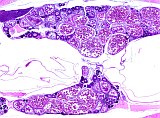
Fig. 1: Ovary, normal 2.5x
|
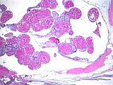
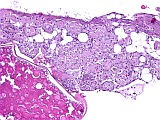
Fig. 2: Atrophic areas of lamellae 2.5x
|
|
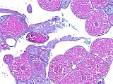
Fig. 3: Atrophic areas of lamellae 5x
|
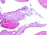
Fig. 4: Atrophic areas of lamellae 10x
|
|
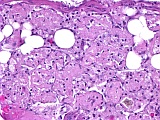
Fig. 5: Atrophic areas of lamellae 20x
|
Fig. 6: Atrophic areas of lamellae 40x
|
Morphologic Description
This ovary has regions with ovigerous lamellae showing normal developing and vitellogenic follicles. However, multifocal segments of the ovigerous lamellae are completely atrophic, with no residual follicular structures. The atrophic segments of lamellae are composed of clusters of macrophages with amorphous eosinophilic material in their cytoplasm. Occasional macrophages filled with yellow-brown lipofuscin-like material are present among the eosinophilic macrophage clusters.
Contributors Comments and Morphologic Diagnoses
Eight contributor posts with comments and/or morphologic diagnoses were submitted. These ranged from a diagnosis of lamellar atrophy with possible residual Leydig cells, possible intersex, ovarian atresia with egg debris, multifocal atrophy/atresia of oocytes, oocyte atresia with interstitial hyperplasia, mesothelial hyperplasia, macrophage aggregates, granulomatous inflammation, increased amount of yolk granules, increased atretic follicles.
I did not observe mesothelial hyperplasia, vacuolated interstitial cells, Sertoli cells, or evidence of intersex. The macrophages present appear to have scavenged previtellogenic atretic oocytes. This pattern differs from the more disorganized granulomatous inflammation occurring in egg-associated inflammation in zebrafish or chronic granulomatous inflammation occurring with piscine mycobacteriosis.
Case Discussion
Zebrafish has become a leading model species for basic biomedical research because of its small size, continuous breeding, short generation time, ease of cloning, fully sequenced genome, and abundance of mutant lines for study of models for human disease. In recent years, several zebrafish mutant lines which are transparent throughout life allow unprecedented in vivo studies of developmental processes, tumor progression, and host-pathogen interactions. Several of the genes mutated in these transparent mutant lines have been associated with ovarian abnormalities when mutated in humans and mice. For example, ovarian hypoplasia and atrophy have occurred in humans and mice with oculocutaneous albinism, vitiligo, Waardenburg syndrome, KIT mutations. Neurogenic neural crest tissue plays an essential role in ovarian development and homeostasis. Thus, mutations in the genes regulating pigment production by neural crest cells can have significant effects on ovarian morphology and function.
Proposed Diagnosis
Severe multifocal locally extensive atrophy of ovigerous lamellae
References
- Antinucci P, Hindges R (2016) A crystal-clear zebrafish for in vivo imaging. Sci Rep 6: 29490.
- Fukamachi S, Asakawa S, Wakamatsu Y, Shimizu N, Mitani H, Shima A (2004) Conserved function of medaka pink-eyed dilution in melanin synthesis and its divergent transcriptional regulation in gonads among vertebrates. Genetics 168(3): 1519-1527.
- Kissel H, Timokhina I, Hardy MP, Rothschild G, Tajima Y, Soares V, Angeles M, Whitlow SR, Manova K, Besmer P (2000) Point mutation in kit receptor tyrosine kinase reveals essential roles for kit signaling in spermatogenesis and oogenesis without affecting other kit responses. EMBO J 19(6): 1312-1326.
- Lang MR, Patterson LB, Gordon TN, Johnson SL, Parichy DM (2009) Basonuclin-2 requirements for zebrafish adult pigment pattern development and female fertility. PLoS Genet 5(11): e1000744.
- McKey J, Bunce C, Batchvarov IS, Ornitz DM, Capel B (2019) Neural crest-derived neurons invade the ovary but not the testis during mouse gonad development. Proc Natl Acad Sci USA 116(12): 5570-5575.
- Meijer AH, Spaink HP (2011) Host-pathogen interactions made transparent with the zebrafish model. Curr Drug Targets 12(7): 1000-1017.
- Parichy DM, Rawls JF, Pratt SJ, Whitfield TT, Johnson SL (1999) Zebrafish sparse corresponds to an orthologue of c-kit and is required for the morphogenesis of a subpopulation of melanocytes, but is not essential for hematopoiesis or primordial germ cell development. Development 126(15): 3425-3436.
- Rawls JF, Johnson SL (2001) Requirements for the kit receptor tyrosine kinase during regeneration of zebrafish fin melanocytes. Development 128(11): 1943-1949.
- Smith ER, Yeasky T, Wei JQ, Miki RA, Cai KQ, Smedberg JL, Yang WL and Xu XX (2012) White spotting variant mouse as an experimental model for ovarian aging and menopausal biology. Menopause 19(5): 588-596.
- Wenner M (2009) The most transparent research. Nat Med 15(10): 1106-1109.
- White RM, Sessa A, Burke C, Bowman T, LeBlanc J, Ceol C, Bourque C, Dovey M, Goessling W, Burns CE, Zon LI (2008) Transparent adult zebrafish as a tool for in vivo transplantation analysis. Cell Stem Cell 2(2): 183-189.
|
|