|
| Guess What! - ESTP Case 17 |
Male, adult New Zealand White rabbit, low dose of a short term toxicity study.
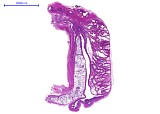
Paramedian section of vesicular gland, prostate, and seminal vesicles.
| Click on the images below for a larger view. |
|
Fig. 1: H&E, x0.2
|
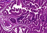
Fig. 2: H&E, x5
|
|
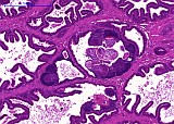
Fig. 3: H&E, x5
|
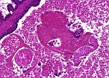
Fig. 4: H&E, x10
|
|
Fig. 5: H&E, x10
|
Morphologic Description
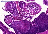
In the prostate and vesicular gland of this New Zealand White rabbit (Oryctolagus cuniculus), multifocal basal cell hyperplasia and squamous metaplasia were observed. The appearance of the changes differed in postate and vesicular gland.
In the prostate (Fig. 2) some alveoli showed an increased proportion of basal cells giving these alveoli a more basophilic appearance.The secretory cells were differentiated similar to normal alveoli in the neighbourhood as columnar glandular epithelia.
In the vesicular gland, nodular hyperplastic foci of basal cells with slight atypia were present which bulged in the direction of the underlying stroma (Fig. 3). Some of the foci showed squamous metaplasia (Fig. 3 and 5). Debris of desquamated epithelia was intermingled with the glandular secretion (Fig. 4).
Proposed Diagnosis
Multifocal basal cell hyperplasia of the prostate and multifocal basal cell hyperplasia with squamous metaplasia of the vesicular gland.
Morphology of the Accessory Male Glands of Rabbits
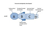
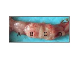
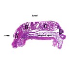
Compared to other species, rabbit accessory male glands are complex and terminology differs among the investigators. Different terms used by Zwicker et al. (1985), Holtz and Foote (1978), Barone et al. 1968). The anatomical terms according to Zwicker et al. (1985) are included in a schematic drawing (Fig. 6) together with names given by other investigators in brackets, except for the paraprostate syn. prostate ventral lobe, which was not mentioned separately in this article. Fig. 7 shows the situation from dorsal and Fig. 8 is a longitudinal paramedian section giving an overview of the glands. The glands with changes are the numbers B (prostate) and C (vesicular gland).
The epithelium of prostate and vesicular gland consists of columnar secretory epithelia and basal cells, the latter being the main proliferative cell type. In addition, endocrine paracrine cells are constituents of the glandular epithelium.
| Click on the images below for a larger view. |
|
Fig. 6: Schematic drawing
|
Fig. 7: Dorsal view
|
|
Fig. 8: Longitudinal paramedian section
|
Discussion
Eight diagnoses were received for this case which were quite uniform: Squamous metaplasia (7), hyperplasia (4), and carcinoma (1). Probably because of the above mentioned difficult anatomy, the localization of changes varied: prostate (5) seminal vesicles (2), vesicular gland (1), no gland mentioned (2).
Histologically, the lesions consisted of basal cells with large pale nuclei. Focal lesions of the vesicular gland, which were generally more nodular, often bulged towards the underlying stroma without any evidence of infiltrative growth. Some of these foci showed squamous metaplasia. Although slight cellular atypia was present, pronounced pleomorphism or increased mitotic rate as indications of possible transition to neoplasia could not be observed.
In the investigation of Zwicker et al. (1985), possible causes of hyperplasia and squamous metaplasia of accessory male glands of rabbits were discussed and reference made to previous investigators who could not link them to inadequate supply with vitamin A or hormonal imbalances. Proliferation and differentiation of the glandular epithelium is influenced by steroid-sensitive cells in the underlying stroma and epithelium. Hormonal imbalances can thus cause diffuse changes of glandular morphology. In rabbits, two weeks of treatment with estrogen induced an increased amount of glandular musculofibrous stroma along with proliferation of the basal cell layer, squamous metaplasia and leukocytic infiltration of the epithelium whereas testosterone treatment promoted differentiation of glandular epithelia (Orgebin-Crist et al. 1983). In this report on induced changes the vesicular gland was not particularly sensitive, and in contrast to the case presented here, other parts of the genital tract exhibited simultaneous stromal or epithelial changes.
Non-productive viral infections of epithelial cells can lead to cellular atypia, hyperplasia and eventually neoplasia. Such findings often arise at mucosal surfaces prone to contact with infectious agents. Although in human prostatic cancer viral sequences have been detected, their significance is still debated. In rabbits, the prostate is not known to be prone to tumor development (Heatley and Smith 2004).
In the human prostate, different types of basal cell hyperplasia including nodular foci and squamous metaplasia are known to occur in up to 10 % of human prostates. They develop either for unknown reasons or associated with inflammation and atrophy (Montironi et al. 2005, 2012). Synonyms for the condition are "fetalization" or "embryonic hyperplasia" referring to the resemblance of nodular basal cell hyperplasia to the morphology of embryonic prostate. An interesting aspect discovered in human prostates is the clonality of neighbouring basal and glandular epithelial cells which implies that a genetic change present in a stem cell is passed down to its decendents (Gaisa et al. 2011). It is likely that this applies also to accessory male glands of other species.
A reason for the frequent spontaneous occurrence of basal cell hyperplasia in rabbits could not be determined. Since reproduction of the animals is unaffected (Zwicker et al. 1985), basal cell hyperplasia in rabbits has no clinical relevance. Nevertheless, the lesions are of interest for the toxicopathologist since they have to be distinguished from hormone-related estrogenic effects.
References
- Barone R, Pavaux C. Blin PC, Cuq P (1973) Atlas d'anatomie du lapin. ISBN 2-225 35 530 7. Masson & Cie, editeurs, Paris, France
- Gaisa NT, Graham TA, McDonald SAC, Poulsom R, Heidenreich A, Jakse G, Knuechel R, Wright NA (2011) Clonal architecture of human prostatic epithelium in benign and malignant conditions. J Pathol 225: 172 - 180
- Heatley JJ and Smith AN (2004) Spontaneous neoplasms of lagomorphs. Vet Clin Exot Anim 7: 561 - 577
- Holtz W, Foote RH (1978) The anatomy of the reproductive system in male Dutch rabbits (Oryctolagus cuniculus) with special emphasis on the accessory sex glands. J Morphol 158: 1 - 20
- Montironi R, Mazzucchelli R, Stramazotti D, Scarpelli M, Lopez Beltran A, Bostwick DG (2005) Basal cell hyperplasia and basal cell carcinoma of the prostate: a comprehensive review and discussion of a case with c-erbB-2 expression. J Clin Pathol 58: 290 - 296
- Montironi R, Scarpelli M, Mazzucchelli R, Cheng L (2012) The spectrum of morphology in non-neoplastic prostate including cancer mimics. Histopathology 60: 41 - 58
- Orgebin-Crist M-C, Eller BC, Danzo BJ (1983) The effects of estradiol, tamoxifen, and testosterone on the weights and histology oft he epididymis and accessory sex organs of sexually immature rabbits. Endocrinology 113: 1703 - 1715
- Zwicker GM, Killinger JM, McConnell RF (1985) Spontaneous vesicular and prostatic gland epithelial squamous metaplasia, hyperplasia. and keratinized nodule formation in rabbits. Toxicol Pathol 13: 222 - 228
|
|