|
| Guess What! - ESTP Case 29 |
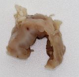
Due to a 5 cm in diameter sized mass at the left side of the neck, a 20-week-old female Wistar (Han) rat was humanely killed.
| Click on the images below for a larger view. |
|
Fig. 1: Makro
|
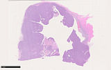
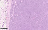
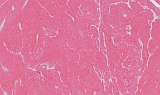
Fig. 2: Overview, H&E
|
|
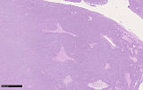
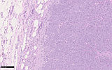
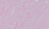
Fig. 3: H&E, 5x
|
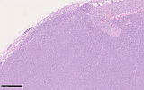
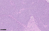
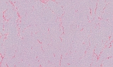
Fig. 4: H&E, 10x
|
|
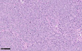
Fig. 5: H&E, 10x
|
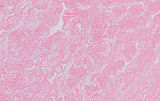
Fig. 6: H&E, 20x
|
|
Fig. 7: H&E, 20x
|
Fig. 8: H&E, 40x
|
Initially, we provided only HE slides and received 7 responses. Five responders suspected a basal cell tumor (four malignant, one benign).
On histology, the mass was unencapsulated and infiltrated the adjacent tissue. The mitotic rate was high with up to 10 mitoses per high-power field. Further, areas of necrosis filled with cellular debris were visible. Therefore, this tumor should be classified as malignant. These features would fit for a basal cell tumor. However, no palisading at the periphery of lobules were apparent. Therefore, the histologic appearance of this tumor does not fit 100% to a basal cell tumor.
One responder suspected a neuroendocrine tumor/carcinoma probably of thyroid or parathyroid origin. A clear neuroendocrine packaging was not observable. The last responder mentioned that this tumor resembles malignant myoepithelioma of mice. All received responds represent valid differential diagnoses. During necropsy we were not able to identify the parotid gland on the left side. Therefore, we also suggested a malignant myoepithelioma. However, this tumor has not been described for rats before.
To further characterize this tumor, we performed immunohistochemistry with antibodies directed against cytokeratin (CK) 5, CK 8, CK 14, pan-cytokeratin, smooth-muscle actin (SMA), vimentin, and podoplanin.
| Click on the images below for a larger view. |
|
Fig. 9: CK5, 10x
|
Fig. 10: CK8, 10x
|
|
Fig. 11: CK14, 10x
|
Fig. 12: PanCK, 10x
|
|
Fig. 13: SMA, 10x
|
Fig. 14: Vimentin, 10x
|
|
Fig. 15: Podoplanin, 10x
|
After providing images of those immunohistochemistry, we received 8. Four responders suggested a malignant myoepithelioma, one a carcinoma, one a basal cell carcinoma of the skin, one a melanoma and one a metastasis of an epitheloid mesothelioma.
The immunhistochemistry showed positivity of the tumor cells for CK 5, CK14, pan-CK, vimentin, podoplanin but negativity for CK 8, SMA.
The tumor in the present case was positive for pan-CK, CK5, CK14 and vimentin, which was also described for myoepitheliomas in other species. Interestingly, this tumor was negative for SMA, which has also been described for myoepitheliomas reported in mice (Sundberg et al., 1991) and man (Jones et al., 1992). Sundberg et al. (1991) suspected that the myoepitheliomas of mice originate from a subset of extraglandular ductal myoepithelial cells being negative for SMA. For pleomorphic adenomas and myoepitheliomas of the salivary gland in humans, it is though that myoepithelial elements might lose their expression of SMA at some stage in tumor development (Jones et al., 1992). Also, the tumor cells were positive for podoplanin, which represents a novel myoepithelial cell marker being positive in human pleomorphic adenoma and other salivary gland tumors with myoepithelial differentiation (Hata et al., 2008; Tsuneki et al., 2013).
The histological appearance of this tumor was consistent with the description of the International Harmonization of Nomenclature and Diagnostic Criteria (INHAND) for myoepitheliomas of mice (Nolte et al., 2016). Due to the positivity for CK5, CK14, pan-CK, vimentin and podoplanin, as well as negativity of SMA, this tumor was diagnosed as a malignant myoepithelioma of the parotid gland arising from the ductal structures. This tumor has already been published (Schaudien et al., 2020).
References
- Hata M, Ueki T, Sato A, Kojima H, Sawa Y (2008) Expression of podoplanin in the mouse salivary glands. Archives of Oral Biology 53: 835-841
- Jones H, Moshtael F, Simpson RHW (1992) Immunoreactivity of a-smooth muscle actin in salivary gland tumours: a comparison with S100 protein. Journal of Clinical Pathology 45: 938-940
- Nolte T, Brander-Weber P, Dangler C, Deschl U, Elwell MR et al. (2016) Non-proliferative and proliferative lesions of the gastrointestinal tract, pancreas and salivary glands of the rat and mouse. Journal of Toxicologic Pathology 29(Suppl. 1): 1S-125S
- Schaudien D, Creutzenberg O, Wagner A, Dahlmann F, Rittinghausen S (2020) Malignant myoepithelioma of the parotid gland in a rat. Journal of Comparative Pathology 176: 162-164
- Sundberg JP, Hanson CA, Roop DR, Brown KS, Bedigian HG (1991) Myoepitheliomas in inbred laboratory mice. Veterinary Pathology 28: 313-323
- Tsuneki M, Maruyama S, Yamazaki M, Essa A, Abe T et al. (2013) Podoplanin is a novel myoepithelial cell marker in pleomorphic adenoma and other salivary gland tumors with myoepithelial differentiation. Virchow’s Archive 462: 297-305
|
|