|
| Guess What! - ESTP Case 2 |
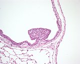
Small exophytic proliferation arising as incidental finding within a bronchiolus of a 16-week-old female Wistar rat (strain: Chbb:Thom SPF) from the low dose group of an inhalation toxicity study. The lesion is composed of central polygonal cells with weakly eosinophilic cytoplasm and large round nuclei and covered by respiratory epithelium.
| Click on the images below for a larger view. |
|
Fig. 1: H&E, x20
|
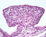
Fig. 2: H&E, x63
|
Morphologic Description
The finding in question was incidentally observed in the bronchiolar epithelium of a 16-week-old female Wistar rat (strain: Chbb:Thom SPF) from the low dose group of an inhalation toxicity study. The lesion is composed of central polygonal cells with weakly eosinophilic cytoplasm and large round nuclei and covered by respiratory epithelium. It consists of an accumulation of neuroendocrine cells located on a stalk of fine fibrovascular tissue.
These neuroendocrine cells were first described as "helle Zellen" by Fröhlich (1949), their presence was later confirmed by Feyrter (1954). Neuroendocrine cells of the airways either occur singly or in clusters known as neuroepithelial bodies. The cells are functionally and structurally related to the cells of the APUD series and is has been postulated that groups of these cells may have sensory receptor sites. For a long time these cells were thought to arise only in the fetal organism but systematic investigations had shown that Feyrter cells occur frequently also in adult rat.
Proposed Diagnosis
Focal epithelial hyperplasia of neuroendocrine cells
Synonym: Aggregation of neuroendocrine (Feyrter) cells
Differential Diagnoses and Discussion
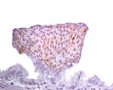
The neuroendocrine origin of the alteration was suggested by about half of the participants. Several colleagues proposed to prove the origin using special staining methods such as NSE, chromogranin, or synaptophysin. The lesion also was expected to be immunoreactive for calcitonin, calcitonin gene-related peptide (CGRP), markers which are described as being specific for neuroendocrine cells. Indeed, the latter staining method gave evidence of the neuroendocrine origin (Figures 3 and 4).
The lesion seems to be too small and too well organized to call it already a benign tumor of neuroendocrine cells. However, a pre-neoplastic lesion (hyperplasia) is not described in the current criteria available for the rat.
The other half of the participants had suggested rather a respiratory epithelial origin of the lesion and names like "bronchiolar polyp", "stromal polyp" or "papilloma composed of non-ciliated bronchial epithelial cells" were given. The neuroepithelial structure might have been overlooked, since in the H&E stain there is no clear tinctorial or morphological difference between respiratory epithelial bronchial cells and neuroendocrine cells. The epithelial stalk moreover gives evidence of a papillomatous appearance.
| Click on the images below for a larger view. |
|
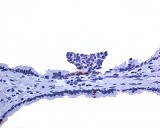
Fig. 3: PCNA, x40
|
Fig. 4: Calcitonin, x63
|
References
- Blair WH (1974) Experimental lung cancer, carcinogenesis and bioassays (Karbe E and Park JF, Eds.) Springer, Berlin New York, pp 199–206
- Dungworth DL, Hahn FF, Hayashi Y, Keenan K, Mohr U, Rittinghausen S, and Schwartz L (1992) International classification of rodent tumours. Part I: The rat (Mohr U, Capen CC, Dungworth DL, Griesemer RA, Ito N and Turusov VS, Eds.) IARC Sci Publ No. 122, Lyon, pp 1–57
- Dungworth DL, Rittinghausen S, Schwartz L, Harkema JR, Hayashi Y, Kittel B, Lewis D, Miller RA, Mohr U, Morgan KT, Rehm S and Slayter MV (2001) International classification of rodent tumors. The mouse (Mohr U, Capen CC, Dungworth DL, Greaves P, Hardisty JF, Hayashi Y, Ito N, Long PH and Krinke G, Eds.) Springer, Berlin, Heidelberg, New York, pp 87–137
- Edmonson NA and Lewis DJ (1980) Distribution and ultrastructural characteristics of Feyrter cells in the rat and hamster airway epithelium. Thorax 35: 371–374
- Ernst H, Heinrichs M, Bargsten G, Kittel B, Rittinghausen S, Dungworth DL and Mohr U (1996) Respiratory system (Jones TC, Dungworth DL and Mohr U, Eds.) 2 edition. Springer, Berlin, Heidelberg, New York, pp 107–116
- Gosney JR and Sissons MCJ (1985) Widespread distribution of bronchopulmonary endocrine cells immunoreactive for calcitonin in the lung of the normal adult rat. Thorax 40: 194–198
- Ito T, Ikemi Y, Kitamura H, Ogawa T and Kanisawa M (1989) Production of bronchial papilloma with calcitonin-like immunoreactivity in rats exposed to urban ambient air. Exp Pathol 36: 89–96
- Ito T, Ikemi Y, Ohmori K, Kitamura H and Kanisawa M (1994) Airway epithelial cell changes in rats exposed to 0.25 ppm ozone for 20 months. Exp Toxic Pathol 46: 1–6
- Ito T, Ohyama K, Kusano T, Usuda Y, Nozawa A, Hayashi H, Ohji H, Kitamura H and Kanisawa M (1997) Pulmonary endocrine cell hyperplasia and papilloma in rats induced by intratracheal injections of extract from particulate air pollutants. Exp Toxic Pathol 49: 65–70
- Shimosegawa T and Said SI (1991) Co-occurrence of immunoreactive calcitonin and calcitonin gene-related peptide in neuroendocrine cells of rat lungs. Cell Tissue Res 264: 555–561
- Sissons MCJ and Gosney JR (1985) Pulmonary endocrine cells immunoreactive for calcitonin in the lungs of fetal and neonatal rats. Thorax 40: 862–865
- Yamamoto K, Nakajima A, Eimoto H, Tsutsumi M, Maruyama H, Denda A, Nii H, Mori Y and Konishi Y (1989) Carcinogenic activity of endogenously synthesized N-nitrosobis (2-hydroxypropyl)amine in rats. Carcinogenesis 10: 1607–1611
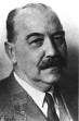 Friedrich Feyrter 1895 - 1973, Pathologist in Vienna, Gdansk, Bratislava, Graz, Vienna and Goettingen Friedrich Feyrter 1895 - 1973, Pathologist in Vienna, Gdansk, Bratislava, Graz, Vienna and Goettingen
|
|