|
| Guess What! - ESTP Case 16 |
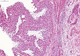
Cynomolgus monkey (Macaca fascicularis); female; approximately 5 years old.
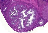
Images show a spontaneous, macroscopically unapparent ovarian lesion.
| Click on the images below for a larger view. |
|
Fig. 1: H&E
|
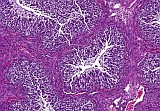
Fig. 2: H&E
|
|
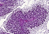
Fig. 3: H&E
|
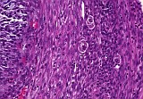
Fig. 4: H&E
|
|
Fig. 5: H&E
|
Fig. 6: H&E
|
Morphologic Description
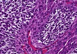
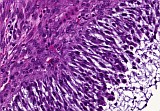
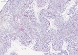
Histopathological examination of case # 16 revealed a unilateral, nodular and well demarcated neoplasm of expansive growth pattern within the ovary of an approximately 5-year-old cynomolgus monkey (Macaca fascicularis). The tumor is characterized by a multicystic, tubulopapillary growth pattern of spindloid to polygonal epithelial cells resembling transitional epithelium. Tumor cells have round to elongated nuclei with multifocal evidence of central longitudinal groove (coffee bean appearance) and pale eosinophilic to mucinous cytoplasm with indistinct cell borders. There is low mitotic activity and mild anisocytosis and anisokaryosis. Within the capsule scattered atretic follicles can be seen.
Proposed Diagnosis
Ovary: Unilateral Brenner tumor
Discussion
Eight diagnoses were received for this case. The diagnosis of Brenner tumor was not included.
Three contributors proposed Sertoli cell tumor/Sertoli cell like tumor as a good differential diagnosis. Another differential diagnosis suggesting sex cord stromal origin was granulosa cell hyperplasia. However, these two entities show slightly different morphological appearance compared to Brunner tumors. Primarily they do resemble transitional epithelium and have therefore been excluded in the present case.
Epithelial neoplasms like papillary cystadenoma, ovarian follicular adenoma and endometrioid adenocarcinoma, as suggested by three other contributors, lack the stromal appearance of tumor cells as seen in this case.
Ectopic uterine tissue can be excluded due to missing morphological resemblance of uterine epithelium.
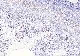
The Brenner tumor of the ovary belongs to the category of transitional cell tumors and represents a relatively rare, mostly benign tumor entity in humans. To our knowledge, it has not yet been described in the Cynomolgus monkey (Macaca fascicularis). It is generally accepted that Brenner tumors are derived from the surface epithelium of the ovary or the pelvic mesothelium through transitional cell metaplasia to form the typical urothelial-like components. Tumor cells usually show positive immunoreactivity for cytokeratin 7, chromogranin (focal) and neuronal marker NSE and are negative for cytokeratin 20 and vimentin. In the present case the immunohistochemical profile differs from Brenner tumors of humans in literature: Tumor cells showed focal positive immunoreactivity for chromogranin (Fig. 7) and pan cytokeratin (pCK) (Fig. 8), whereas reaction for CK 7 and 20 and NSE was negative. Instead, cells were broadly positive for vimentin (Fig. 9). Nevertheless, due to broad concordance of morphologic features, the present case has been classified as a Brenner tumor.
| Click on the images below for a larger view. |
|
Fig. 7: chromogranin, x9
|
Fig. 8: pCK, x20
|
|
Fig. 9: vimentin, x10
|
References
- Bora T, Mahanta RK, Bora BD, Saikia S (2011) Brenner tumor of ovary: An incidental finding. J Midlife Health 2(1): 40–41
- Scully RE (1977) Ovarian tumors. A review. Am J Pathol 87(3): 686-720
- Seidman JD, Khedmati F (2008) Exploring the Histogenesis of Ovarian Mucinous and Transitional Cell (Brenner) Neoplasms and Their Relationship With Walthard Cell Nests: A Study of 120 Tumors 132 (11): 1753-1760
- Ziadi S, Trimeche M, Hammedi F, Hidar S, Sriha B, Mestiri S, Korbi S (2010) Bilateral proliferating Brenner tumor of the ovary associated with recurrent urothelial carcinoma of the urinary bladder. N Am J Med Sci 2(1): 39-41
|
|