|
| Guess What! - ESTP Case 1 |
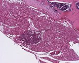
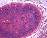
The lesion was observed as incidental finding in a 10-week-old female Wistar rat from a subchronic toxicity study. It was localized within the brown fat tissue adjacent to the thoracic vertebral column and appeared to be associated with a larger venule. The animal did not show any macroscopic or further unusual histopathological findings.
| Click on the images below for a larger view. |
|
Fig. 1: H&E, x10
|
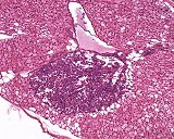
Fig. 2: H&E, x20
|
|
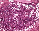
Fig. 3: H&E, x40
|
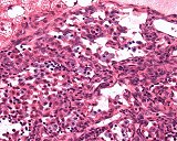
Fig. 4: H&E, x60
|
Morphologic Description
The alteration is surrounded by a thin capsule and consists of several vascular channels which are partly filled with erythrocytes and diffusely distributed lymphocytes. The latter do not show any follicular arrangement. The endothelial lining of the vascular structures seems to be regular and does not exhibit any hypertrophic cells or enlarged nuclei. Single or small clusters of mast cells are loosely distributed in the interstitial connective tissue.
Proposed Diagnosis
Angiomatous hamartoma
Differential Diagnoses and Discussion
"Focal angiomatous hyperplasia" could be considered as an alternative diagnosis. However, the presence of lymphocytes and mast cells is not a characteristic feature of this potentially pre-neoplastic lesion. Also, hemangioma/-sarcoma which occasionally arises in the subcutaneous adipose tissue, does not reveal such associated cell populations. Thus, by its morphologic features the presented alteration rather resembles the structures of a lymph node, in which intermingled mast cells are frequently observed.
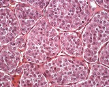
Consequently, one of the contributing participants suggested the presence of a "hemolymphatic node". Such hemal or splenoid nodes, as they are also named, occur physiologically in ruminants in the vicinity of larger vessels, especially in cattle. They have been found also in primates and horses. With respect to their structure and function they represent an intermediate between lymph node and spleen. The hemal node is characterized by a large, endothelium-lined and blood-filled subcapsular sinus that communicates with a network of deep blood sinuses and lymphatic follicles and cords (Fig. 5). The cords are composed of loosely-arranged lymphocytes enmeshed in a reticular network. The lymphatic follicles may have typical germinal centers surrounded by mature lymphocytes (Fig. 6). However, the typical lymphofollicular structures and/or a regular sinus system are lacking in the presented case.
Another contributor suggested the diagnosis "chemodectoma". This neuroectodermal neoplastic lesion (non-chromaffine paraganglioma) shows pathognomonic spherical structures (so-called "Zellballen") which are composed of polygonal chief cells with prominent eosinophilic or finely granular cytoplasm. The "Zellballen" are surrounded by dense bundles of reticulin fibers which, especially in reticulin stains, produce the characteristic nesting (alveolar) pattern of the tumor. Comparable structures, however, are missing in the presented case, where a vascular pattern predominates (Fig. 7 and 8).
| Click on the images below for a larger view. |
|
Fig. 5: H&E
|
Fig. 6: H&E
|
|
Fig. 7: H&E x20
|
Fig. 8: H&E x40
|
|
|