|
| Guess What! - ESTP Case 22 |
Animal details:
Male rat of a juvenile toxicity study (high dose groups) which survived the scheduled 8 week treatment period.
The animal was treated daily by oral gavage starting on post natal day 4.
So the age of the animal at necropsy was 8.5 weeks.
Necropsy findings:
At macroscopic examination a dark red nodule of 9x19 mm in diameter was present on the skull.
There were no other macroscopic findings.
A slide from the nodule was prepared after decalcification of the tissue and stained with H&E.
Morphologic Description
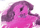
A male Wistar (Han) rat from a juvenile oral gavage toxicity study was presented at necropsy with a dark red nodule of 9x19 mm in the subcutaneous connective tissue of the skullcap. The overlying skin was intact and was not attached to the nodule.
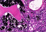
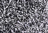
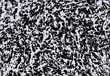
At microscopic examination, the nodule consisted of proliferated osseous tissue with bone marrow, which on the dorsal and lateral surface was covered by dense aggregates of heavily pigmented macrophages intermixed with fibrous tissue. The macrophages were characterized by a large amount of cytoplasm filled with fine to large black pigmented granules. There was no invasion or destruction of adjacent tissue (fig.1, 3).
| Click on the images below for a larger view. |
|
Fig. 1: H&E, x2.4
|
Fig. 2: H&E, x10
|
|
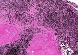
Fig. 3: H&E, x13
|
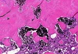
Fig. 4: H&E, x40
|
|
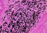
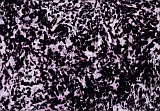
Fig. 5: H&E, x53
|
From the dorsal surface the pigmented macrophages and fibrous tissue showed a linear penetration of the osseous nodule (fig. 2, 4). Free black pigment was also visible in the bone marrow (fig. 5).
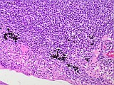
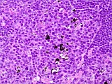
The mandibular lymph nodes showed a slight amount of inert black pigment, resembling tattoo pigment, which can occasionally be seen in draining lymph nodes of tattooed animals in toxicity studies (fig. 6, 7).
| Click on the images below for a larger view. |
|
Fig. 6: H&E, x20
|
Fig. 7: H&E, x40
|
|
Fig. 8: CD68/ED1, x40
|
Fig. 9: S100, x40
|
|
Fig. 10: H2O2, x40
|
Additional investigations were performed to further identify the nodule:
- Immunohistochemistry for CD68/ED1 (macrophage marker - chromogen was Fast Red): Nearly all pigmented cells of the lesion were positive (Fig. 8).
- Immunohistochemistry stain for S100 (marker for melanocytic differentiation - chromogen was Fast Red): Only very few cells in the lesion were positive (Fig. 9).
- Bleaching of a section of the nodule with H2O2 (bleaching possible in case of melanin): No bleaching possible (Fig. 10).
Discussion
Ten contributions were received for this case.
(Malignant) melanoma was suggested by eight of the contributors.
Malignant melanotic melanomas are exceedingly rare incidental tumors in rats and have to my knowledge only been reported in pigmented strains. These tumors arise from neuroectodermal cells (melanocytes) which have migrated into the epidermis and epidermal adnexa. Predilection sites are skin of the external ear, genitalia, tail and eye (Zurcher and Roholl, 1989). Similar to the current case, they may show a dense nodular proliferation in the dermis without association to the dermis. The tumor cells are polygonal, epitheloid or spindle-shaped with a varying degree of pigmentation. Melanin-containing macrophages often occur (Bader et al., 1993; Mecklenburg et al., 2013). In contrast to the nodule of the current case, cellular and nuclear pleomorphism are moderate and melanomas in general show invasive growth and destroy pre-existent adjacent structures.
S100 has been shown to be one of the most sensitive markers for melanocytic differentiation, being diffusely positive in all benign, primary and secondary malignant melanomas (de Vries et al., 2001). In veterinary medicine S100 stain is used as a diagnositic criterium for melanomas in several species including, horse, cattle, dog, mouse, guinea pig and rat (Javanbakht et al., 2014). S100 stain of the current nodule showed only very few positive cells.
Heavily pigmented melanomas can be bleached using H2O2 (Lawrence Li et al., 1999; Javanbakht et al., 2014). The H2O2 method on a section of the nodule of the current case didn't result in any bleaching of the black pigment.
Other proposed diagnoses were neurocutaneous melanosis (one contributor) and a skull fracture with reactive melanocytic fibrosis (one contributor).
Neurocutaneous melanosis is a rare, non-familial, congenital syndrome characterized by multiple pigmented nevi, characterized by proliferation of melanin-producing cells in the skin and brain in newborn babies. There is one publication of this lesion in Cynomolgus Macaques (Chen et al., 2009). It has to my knowledge not been reported in other species. The present lesion in the male Wistar (Han) rat didn't contain melanin pigment, as evidenced by the inability to bleach the nodule with H2O2 and the negative S100 stain.
A skull fracture with reactive melanocytic fibrosis should be considered a differential diagnosis. The lesion with proliferated bone and fibrous tissue is similar to what can be seen after a fracture. However, what remains unclear with this suggestion is the origin of the black pigment. The fibrotic tissue doesn't contain melanin as evidenced by the absence of S100 positive staining properties and the inability to bleach with H2O2.
Proposed Diagnosis
Granulomatous inflammatory process with abundant black (Indian ink) pigment
For this lesion the diagnosis was considered to be a granulomatous inflammatory process with abundant black pigment. The complete history of this animal was investigated and this showed that the juvenile rats in the current study were identified with a subcutaneous injection with Indian ink on postnatal day 1 or 2. The male rat presented in this case received a subcutaneous injection in the neck region. During this procedure the Indian ink was probably accidently injected (partly) in the soft neonatal skull. This likely resulted in reactive proliferation and an inflammatory process with macrophages containing black (Indian ink) pigment. The linear macrophage infiltrate in the bone might mark the needle track formed by the injection with Indian ink.
References
- Bader R, Gembardt C, Kaufmann W, Kuettler K, Mann PC, van Zwieten MJ and Zurcher C (1993) 5. Integumentary System. In: Mohr U, Capen CC, Dungworth DL, Griesemer RA, Ito N, Turusov VS (eds) International classification of rodent tumours, Part I: The Rat. IARC Scientific Publications No. 122, Lyon, pp 1–21.
- Chen Y, Deng W, Zhu H, Li J, Xu Y, Dai X, Jia C, Kong Q, Huang L, Liu Y, Ma C, Xiao C, Liu Y, Li Q, Bezard E, Qin C. (2009) The pathologic features of neurocutaneous melanosis in a Cynomolgus Macaque. Vet Pathol 46: 773-775.
- de Vries TJ, Smeets M, de Graaf R, Hou-Jensen K, Bröcker EB, Renard N, Eggermont AM, van Muijen GN, Ruiter DJ (2001) Expression of gp100, MART-1, tyrosinase, and S100 in paraffin-embedded primary melanomas and locoregional, lymph node, and visceral metastases: implications for diagnosis and immunotherapy. A study conducted by the EORTC Melanoma Cooperative Group. J Pathol 193(1): 13-20.
- Javanbakht J, Sasani F, Adibhashemi F, Hemmati S (2014) Comparative histopathological diagnosis of cutaneous melanoma by H&E, special staining and immunohistochemical methods against cutaneous squamous cell carcimoma in horse and bovine. J Bioanal Biomed 6(4): 19-23.
- Lawrence Li L, Crotty KA, Kril JJ, Palmer AA, McCarthy SW (1999) Method of melanin bleaching in MIB1-Ki67 immunostaining of pigmented lesions: A quantitative evaluation in malignant melanomas. Histochem J 31: 237-240.
- Mecklenburg L, Kusewitt D, Kolly C, Treumann S, Terence Adams E, Diegel K, Yamate J, Kaufmann W, Müller S, Danilenko D, Bradley A (2013) Proliferative and non-proliferative lesions of the rat and mouse integument. J Toxicol Pathol 26 (3 Suppl): 27S–57S.
- Zurcher C, Roholl PJM (1989) Melanocytic tumors, rat. In: Jones TC, Mohr U, Hunt RD (eds) Monographs on Pathology of Laboratory Animals, Integument and Mammary Glands. Springer, Berlin, pp 76-86.
|
|